

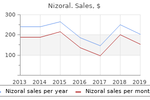
Nizoral
Order nizoral 200mg free shipping
Like a pacemaker system, a pacing cardioverter defibrillator system additionally features a pulse generator and electrodes, although pacing cardioverter-defibrillators might require a number of leads, even when solely a single chamber is being paced. A pacing cardioverter-defibrillator system could also be inserted in a single chamber (pacing the ventricle) or in dual chambers (pacing the atrium and ventricle). These units use a mix of antitachycardia pacing, low vitality cardioversion or defibrillating shocks to deal with ventricular tachycardia or ventricular fibrillation. Pacing cardioverter-defibrillator pulse turbines could also be implanted in a subcutaneous infraclavicular pocket or in an abdominal pocket. Removal of a pacing cardioverter-defibrillator pulse generator requires opening of the present subcutaneous pocket and disconnection of the heart beat generator from its electrode(s). The electrodes (leads) of a pacing cardioverter-defibrillator system are positioned in the heart through the venous system (transvenously), in most circumstances. In certain circumstances, an additional electrode could also be required to realize pacing of the left ventricle (bi-ventricular pacing). In this event, transvenous (cardiac vein) placement of the electrode ought to be individually reported using code 33224 or 33225. Epicardial placement of the electrode ought to be individually reported using 3320233203. Electrode positioning on the epicardial surface of the guts requires thoracotomy, or thoracoscopic placement of the leads. Removal of electrode(s) might first be attempted by transvenous extraction (code 33244). However, if transvenous extraction is unsuccessful, a thoracotomy could also be required to remove the electrodes (code 33243). Use codes 33212, 33213, 33240 as acceptable along with the thoracotomy or endoscopic epicardial lead placement codes to report the insertion of the generator if done by the same physician throughout the same session. Replacement of a pulse generator ought to be reported with a code for removal of the heart beat generator and one other code for insertion of a pulse generator. Repositioning of a pacemaker electrode, pacing cardioverter-defibrillator electrode(s), or a left ventricular pacing electrode is reported using 33215 or 33226, as acceptable. Replacement of a pacemaker electrode, pacing cardioverter-defibrillator electrode(s), of a left ventricular pacing electrode is reported using 33206-33208, 33210-33213, or 33224, as acceptable. Version 2020 Page a hundred and one of 258 Physician Procedure Codes, Section 5 Surgery 33202 Insertion of epicardial electrode(s); open incision (eg, thoracotomy, median sternotomy, subxiphoid approach) 33203 endoscopic approach (eg, thoracoscopy, pericardioscopy) (When epicardial lead placement is carried out by the same physician at the similar session as insertion of the generator, report 33202, 33203 along side 33212, 33213, as acceptable) 33206 Insertion of recent or alternative of everlasting pacemaker with transvenous electrode(s); atrial 33207 ventricular 33208 atrial and ventricular (Codes 33206-33208 embody subcutaneous insertion of the heart beat generator and transvenous placement of electrode(s)) 33210 Insertion or alternative of momentary transvenous single chamber cardiac electrode or pacemaker catheter (separate process) 33211 Insertion or alternative of momentary transvenous dual chamber pacing electrodes (separate process) 33212 Insertion of pacemaker pulse generator solely; with current single lead 33213 with current dual leads (When epicardial lead placement is carried out with insertion of generator, report 33202, 33203 along side 33212, 33213) 33214 Upgrade of implanted pacemaker system, conversion of single chamber system to dual chamber system (consists of removal of previously placed pulse generator, testing of current lead, insertion of recent lead, insertion of recent pulse generator) (Do not report 33214 along side 33227-33229) 33215 Repositioning of previously implanted transvenous pacemaker or implantable defibrillator (proper atrial or proper ventricular) electrode 33216 Insertion of a single transvenous electrode, everlasting pacemaker or implantable defibrillator 33217 Insertion of two transvenous electrodes, everlasting pacemaker or implantable defibrillator 33218 Repair of single transvenous electrode, everlasting pacemaker or implantable defibrillator 33220 Repair of two transvenous electrodes for everlasting pacemaker or implantable defibrillator 33221 Insertion of pacemaker pulse generator solely; with current a number of leads 33222 Relocation of pores and skin pocket for pacemaker 33223 Relocation of pores and skin pocket for implantable defibrillator 33224 Insertion of pacing electrode, cardiac venous system, for left ventricular pacing, with attachment to previously placed pacemaker or implantable defibrillator pulse generator (including revision of pocket, removal, insertion, and/or alternative of current generator) (When epicardial electrode placement is carried out, report 33224 along side 33202, 33203) 33225 Insertion of pacing electrode, cardiac venous system, for left ventricular pacing, at time of insertion of implantable defibrillator or pacemaker pulse generator (eg, for upgrade to dual chamber system) (List individually along with primary process) (Use 33225 along side 33206, 33207, 33208, 33212, 33213, 33214, 33216, 33217, 33221,33223, 33228, 33229, 33230, 33231, 33233, 33234, 33235, 33240, 33249, 33263, 33264) Version 2020 Page 102 of 258 Physician Procedure Codes, Section 5 Surgery 33226 Repositioning of previously implanted cardiac venous system (left ventricular) electrode (including removal, insertion and/or alternative of current generator) 33227 Removal of everlasting pacemaker pulse generator with alternative of pacemaker pulse generator; single lead system 33228 dual lead system 33229 a number of lead system (Do not report 33227-33229 along side 33233) 33230 Insertion of implantable defibrillator pulse generator with current dual leads 33231 with current a number of leads (Do not report 33230, 33231, 33240 along side 33241 for removal and alternative of the pacing cardioverter-defibrillator pulse generator. Tissue ablation, disruption and reconstruction could be completed by many strategies including surgical incision or via the usage of a wide range of vitality sources (eg, radiofrequency, cryotherapy, Version 2020 Page 103 of 258 Physician Procedure Codes, Section 5 Surgery microwave, ultrasound, laser). Additional ablation of atrial tissue to remove sustained supraventricular dysrhythmias. This must embody operative ablation that includes both the proper atrium, the atrial septum, or left atrium in continuity with the atrioventricular annulus. A subcutaneous cardiac rhythm monitor is placed using a small parasternal incision followed by insertion of the monitor right into a small subcutaneous prepectoral pocket, followed by closure of the incision. Version 2020 Page 107 of 258 Physician Procedure Codes, Section 5 Surgery See 33517-33523 and 33533-33536 for reporting combined arterial-venous grafts. To report harvesting of an higher extremity vein, use 35500 along with the bypass process. When surgical assistant performs graft procurement, add modifier �80 to 33510-33516. To report harvesting of an higher extremity artery, use 35600 along with the bypass process. To report harvesting of a femoropopliteal vein phase, report 35572 along with the bypass process. When surgical assistant performs arterial and/or venous graft procurement, add modifier 80 to 33517-33523, 33533-33536, as acceptable. The codes embody the usage of the internal mammary artery, gastroepiploic artery, epigastric artery, radial artery, and arterial conduits procured from different sites. To report harvesting of an higher extremity vein, use 33500 along with the bypass process. To report harvesting of a femoropopliteal vein phase, report 33572 along with the bypass process. When surgical assistant performs arterial and/or venous graft procurement, add modifier 80 to 33517-33523, 33533-33536 as acceptable.
Buy cheap nizoral 200 mg online
Bonilla-Musoles F, Raga F, Osborne N, Blanes J: the usage of three-dimensional (3D) ultrasound for the study of regular and pathological morphology, of the human embryo and fetus: preliminary report. Fujiwaki R, Hata T, Hata K, Kitao M: Intrauterine sonographic assessments of embryonic growth. Merz E: Three-dimensional ultrasound within the analysis of fetal anatomy fetal malformations. Steiner H, Staudach A, Spitzer D, Schaffer H: Three-dimensional ultrasound in obstetrics and gynecology: method, prospects and limitations. The chorionic vasculature is studied, and notes on aberrancies ought to be recorded. The twine and membranes are trimmed from the placenta earlier than its weight is decided. The parenchyma of the placenta is examined in �bread loaf� sections to establish irregularities. A membrane roll is made, which is derived from spiral rotation of the membranes with the point of rupture on the center. Sections to be microscopically examined include a cross part of the twine, the membrane roll, and three sections from the parenchyma including central and peripheral areas of the placental disc. Small placentas are seen in preeclampsia, low delivery weight, and accelerated villous maturation. Large placentas are seen with villous edema, extreme maternal anemia, fetal anemia, syphilis, large intervillous thrombi, maternal diabetes, subchorionic thrombosis,toxoplasmosis,congenitalfetalnephrosis,idiopathicfetalhydrops, and a number of placental chorangiomas (Table 5. Severe anemia of the fetus is seen with large quantities of fetal�maternal hemorrhage. Congenital infections, such as parvovirus B19, will produce anemia and is seen as pallor within the placenta. In placenta membranacea, the complete chorion laeve persists and covers the membranes. Theseabnormalplacentas are sometimes associated with placenta accreta, second-trimester bleeding, and fetal and neonatal problems. The placenta has penetrated into the muscularis of the has persisted and covers the membranes on left. Retroplacental hemorrhage on materpenetrated by way of the total thickness of the nal surface leading to abruption. Placental findings include atrophy or infarction of villi near the cervical os as a result of many such pregnancies are marked by secondand third-trimester bleeding, retroplacental hemorrhage, or hemosiderin deposition within the membranes. Preterm delivery, in addition to chronic bleeding and associated maternal and fetal anemia, increase the danger of hypoxia. There can be a positive correlation between cigarette smoking and the discovering of placenta previa. Findings within the placenta include attribute intervillous hemorrhage and associated early modifications of infarction (Figures 5. Thrombotic material is often found adherent to the base of the placenta that will depress its surface. A Causes of placental abruption include chorioamnionitis, hypertensive illness, infiammation of the decidual vasculature, and trauma. A long umbilical twine, congenital malformations, preeclampsia, and smoking throughout being pregnant end in an elevated threat of placental abruption. Intervillous thrombi are answerable for fetal�maternal hemorrhage and associated fetal anemia (Figure 5. Maternal systemic lupus erythematosus has circulating antiphospholipid and anticardiolipin antibodies and is associated with second trimester abortions. The placenta shows much intervillous fibrin and immunoglobulin deposits of principally IgG and IgM. In hypertensive pregnancies, attribute decidual vascular problems diminish blood fiow to the villous parenchyma and end in infarction. Abruption, an infection, immunologic problems, smoking, and possibly cocaine all correlate with villous ischemia and infarction.
Syndromes
- Itchy nose, mouth, eyes, throat, skin, or any area
- Leukemia
- Have a new rash or bruises appear
- The skin might be cracking, peeling, or blotchy, but this should improve over time.
- If you had a PSA test in the past and how much and how fast your PSA levels have changed
- Gas
- Pneumonia
- Corrosives, such as sodium hydroxide (lye)
Discount nizoral 200 mg with visa
The intraocular pressures (<14 mmHg) and corneal thicknesses (<588 �m) of all 4 eyes have been inside normal vary. Both subjects� eyes revealed open angles on gonioscopy without peripheral anterior synechiae. H, Corneal guttae appeared as dark bulges in between the endothelial cells, with a hyperrefective dot in the middle. This diversity is enhanced by 4 attribute manifestations of corneal aging: stromal microdots, folds in the posterior stroma, opacifcation of Descemet�s membrane, and corneal guttae. These common degenerative processes have been studied in clinically normal corneas and will, in a minority of instances, progress into cornea farinata, posterior crocodile shagreen, or Fuchs� endothelial dystrophy. We discuss, from epithelium to endothelium, the eight most common morphologic variants of the diferent corneal layers that have been noticed in our research, complemented by a novel phenotype of corneal endothelium. Efects of aging on the normal corneal morphology as noticed by in vivo confocal microscopy Age class, years 20-29 30-39 40-forty nine 50-59 60-69 70-79 Eyes, n 60 60 60 60 forty two 18 P or (ninety five% cI) Bright superfcial 9 (15) 6 (10) 6 (10) 4 (7) 10 (24) three (17) 0. These bright cells have been advised to indicate a loss of contact in the process of desquamation. Therefore, appearance of bright superfcial epithelial cells is a phenomenon typically encountered in ophthalmic practice. Dendritic cells serve as a major link between the innate and adaptive immune methods by capturing and processing antigens and presenting them to lymphocytes. A, His proper eye confirmed a dark-mild reversal of the endothelial cells and precipitated granules (arrow) on a fats endothelial layer. The middle of practically all endothelial cells confirmed a variable-sized black discoloration. Nevertheless, we demonstrated that dendriform cell prevalence was constant over age. Because adjustments in the density and morphology of dendriform cells may refect the immune standing of the cornea,5,7 future analysis ought to concentrate on the appearance of dendriform cells during observe-up of chronic infammatory processes, similar to herpetic stromal keratitis. Epithelial basement membrane dystrophy is characterised by maps, dots, fngerprint traces, and blebs as noticed by slit-lamp examination,25�27 whereas the identical alterations How normal is the transparent corneafi Nerves in the mid-stroma have been reported as thick, straight nerves with a mainly dichotomous branching pattern. However, similar tortuous stromal nerves with a beaded appearance have been identifed in the normal corneas of forty three% of healthy subjects. By using immunohistochemical staining, tortuous nerves have been proven to originate from the straight mid-stromal nerves to kind the mid-stromal and subepithelial nerve plexus, which each show a patchy distribution in the central cornea and enhance in density toward the periphery. Therefore, the tortuous nerves are thought to characterize a functionally distinct nerve inhabitants that may provide epithelial reinnervation after corneal harm or possess specifc sensory or trophic capabilities. These small particles have been postulated to characterize dysgenic or apoptotic mobile remnants, similar to lipofuscin granules, which constitute a degenerative process also referred to as �cornea farinata. This enhance of microdots probably brought on backscatter in the stroma to increase in subjects aged fi50 years, as we found in a parallel research. Folds in the posterior stroma have been reported in 10%, 18%, and 29% of the inhabitants in the sixth, seventh, and eighth a long time of life, respectively. Applanation of the cornea has been proven to induce the usually invisible anterior corneal mosaic, posterior stromal bands, and posterior floor ridges. Posterior crocodile shagreen ought to be distinguished from the presence of stromal edema, which also will increase the visibility and severity of the folds60 of the posterior corneal mosaic. In normal corneas, Descemet�s membrane contains an anterior banded layer and a posterior nonbanded layer. Corneal guttae are focal excrescences of Descemet�s membrane that characterize irregular collagen deposition by distressed endothelial cells. This degenerative process is more generally noticed in women67 and has a lower prevalence in Asians. Efects of aging on corneal morphology forty seven a long time, confrming, in vivo, the results of Kaufman et al,72 who reported corneal guttae in 20% of all donor corneas aged fi50 years. The novel phenotype of corneal endothelium, which was noticed in each eyes of two healthy subjects, completely illustrates the morphologic diversity of the transparent cornea. A, Fuchs� endothelial dystrophy is characterised by corneal guttae, noticed as bright spots surrounded by a dark halo interspersed between enlarged endothelial cells.
Buy nizoral 200mg without a prescription
The intent of this document is to teach and help these caring for youngsters with cleft lip and palate. It supplies a framework for a constant approach to administration of those youngsters. This document was created by the cleft lip/palate consensus team, made up of major care physicians, specialty providers, regional cleft lip/palate team coordinators, mother and father and third-get together payers. Central to those paperwork, that are summarized within the appendix, is the precept that sufferers with cleft lip/palate are greatest cared for by an interdisciplinary team of specialists with experience on this feld. The goals of treatment for the child with a cleft lip/palate are: � Repair the birth defect (lip, palate, alveolar ridge, nostril) � Achieve normal speech, language and listening to � Achieve functional dental occlusion and good dental health � Optimize psychosocial and developmental outcomes � Minimize costs of treatment � Facilitate ethically sound, household-centered, culturally sensitive care Seven key themes are necessary for attaining these goals: � Early evaluation and intervention is crucial and should begin within the prenatal or new child interval with referral to a Cleft Lip/Palate Team. Tables on pages 17 and 18 highlight key interventions by self-discipline and age group, respectively. These are explained more absolutely within the body of the document and within the sections that follow. A glossary of phrases, description of cleft varieties, and resource information with a list of cleft lip/palate groups in Washington state are also included. The following pages record issues and interventions for the child with a cleft lip/palate. Most of the interventions listed are supplied by specialists on the cleft lip/palate groups. The services that end result should be closely coordinated with the treatment plans of the patient�s cleft lip/palate team. In addition, cleft lip/palate groups vary in both the disciplines taking part and the interventions supplied. As the interventions listed are essentially brief, appendices have been included to provide extra info in most of the key areas. Referrals to those services can be facilitated by any Children with Special Health Care Needs Coordinator at local public health departments or by the cleft lip/palate team coordinator. Conversely, some sufferers may require interventions not talked about in these suggestions. Each patient�s care plan must be individualized considering medical wants, psychosocial and cultural variables, and resources obtainable in every community. Communication between the community provider and the cleft lip/palate team members is important for creating and implementing these care plans. Some have both cleft lip and palate; some have solely a cleft of the lip (also referred to as the first palate); others have solely a cleft of the palate (also referred to as the secondary palate). With any type of cleft lip, there may be further, lacking or poorly shaped tooth within the space(s) of the cleft. As the child grows and earlier than the palate is repaired, this opening also permits an excessive amount of airfow via the nostril, inflicting hypernasal speech. With improvements in ultrasound technology, the prenatal prognosis of isolated cleft lip is increasingly widespread. A latest meta-evaluation revealed that the majority research reported detection rates between 9-50%, indicating a substantial proportion of diagnoses 5 of cleft lip and palate are nonetheless missed. In the United Kingdom, routine views of the face and lips were added to antenatal ultrasound pointers in 2000 and detection rates of cleft lip in low threat populations elevated from 16-33% to 75% with 2D ultrasound between 18-23 6 weeks gestation. However, if an anticipating mother is being scanned early, for example, if amniocentesis is being thought of (usually at 15-17 weeks), an additional later scan can be performed if there are concerns about a potential cleft. Multiple factors afect ultrasound high quality: fetal place on the time of ultrasound, the mother�s weight, the quantity of amniotic fuid, the type of ultrasound 12 machine, and experience of the sonographer. Once a cleft lip and/or palate is identifed, the household must be referred for genetic counseling to discuss choices for added testing, together with amniocentesis. This ought to include info on any teratogenic exposures, maternal health points, and the presence of relations with clefts or other congenital variations, developmental issues and genetic syndromes. If prenatal ultrasounds and tests reveal anomalies along with the cleft, the potential for a syndrome or chromosome diference is more likely. Even if genetic tests are unfavorable, mother and father must be informed that an correct prognosis and full discussion of prognosis and recurrence risks can solely happen after the infant is born. The experience of learning about a cleft in an unborn child will have associated emotions and reactions that may be diferent for every particular person. The strategy of restoration guides a household right into a mode of problem fixing, and opens up opportunities for learning about cleft care 15 and their child�s wants.
200mg nizoral with visa
Peripheral Ulcerative Keratitis (Marginal Keratitis in Autoimmune Disease) (Figure 6�9) Figure 6�9. Three hundred sixty levels of peripheral ulcerative keratitis in a affected person with rheumatoid arthritis. The peripheral cornea receives its nourishment from the aqueous humor, the limbal capillaries, and the tear movie. It is contiguous with the subconjunctival lymphoid tissue and the lymphatic arcades at the limbus. The perilimbal conjunctiva seems to play an essential position within the pathogenesis of corneal lesions that come up each from local ocular illness and from systemic issues, notably those of autoimmune origin. There is a putting similarity between the limbal capillary network and the renal glomerular capillary network. On the endothelial basement membranes of the capillaries of each networks, immune complexes are deposited and immunologic illness outcomes. Thus, the peripheral cornea often participates in such autoimmune ailments as rheumatoid arthritis, polyarteritis nodosa, systemic lupus erythematosus, scleroderma, granulomatosis with polyangiitis (Wegener�s granulomatosis), ulcerative colitis, Crohn�s illness, and relapsing polychondritis. The corneal adjustments are secondary to scleral inflammation, with or with out scleral vascular closure (see Chapter 7). The medical indicators embody vascularization, infiltration and opacification, and peripheral guttering that may progress to perforation. Treatment is directed toward management of the related systemic illness; topical remedy normally is ineffective, and systemic use of potent immunosuppressants often is required. Corneal perforation could require cyanoacrylate glue (Figure 6�5), lamellar patch 298 grafting, or full-thickness keratoplasty. Vitamin a Deficiency the everyday corneal ulcer related to avitaminosis A is centrally positioned and bilateral, gray, and indolent, with a definite lack of corneal luster within the surrounding area. The epithelium of the conjunctiva is keratinized, as evidenced by the presence of a Bitot�s spot. This is a foamy, wedge-formed area within the conjunctiva, normally on the temporal facet, with the base of the wedge at the limbus and the apex extending toward the lateral canthus. Within the triangle, the conjunctiva is furrowed concentrically with the limbus, and dry flaky materials could be seen falling from the area into the inferior cul-de-sac. A stained conjunctival scraping from a Bitot�s spot will present keratinized epithelial cells. Avitaminosis A corneal ulceration normally outcomes from dietary lack of vitamin A or impaired absorption from the gastrointestinal tract. Lack of vitamin A causes a generalized keratinization of the epithelium throughout the physique. Since the epithelium of the air passages is affected, many sufferers, if not handled, will die of pneumonia. Mild vitamin A deficiency ought to be handled in adults with a dose of 30,000 U/d for 1 week. Neurotrophic Keratitis Trigeminal nerve dysfunction, due to trauma, surgical procedure, tumor, inflammation, or some other trigger, could end in corneal anesthesia with lack of the blink reflex, one of many cornea�s defense mechanisms, as well as lack of trophic components essential for epithelial function. In the absence of corneal sensation, even a extreme keratitis could produce little discomfort. Patients must be warned to look out for redness of the eye, decreased vision, corneal abnormality, or elevated conjunctival discharge and to seek ophthalmic care as soon as any of these develop. Keeping the cornea moist with artificial tears and lubricant ointments could assist to protect it. The most effective administration is to keep the eye closed by cautious horizontal taping of the eyelids, by tarsorrhaphy, or via ptosis induced with botulinum toxin. Topical nerve progress factor drops can be utilized to advertise the therapeutic of epithelial defects, and autologous serum drops could also be helpful in long-term floor maintenance. Examples embody proptosis from any trigger, ectropion, floppy lid syndrome, the absence of a part of an eyelid as a result of trauma, and incapability to shut the lids, as in Bell�s palsy. The two components at work are the drying of the cornea and its publicity to minor trauma. The uncovered cornea is particularly subject to drying throughout sleeping hours, and swim goggles could also be useful at night time. If an ulcer develops, it normally follows minor trauma and occurs within the inferior third of the cornea.
Cheap nizoral 200 mg visa
The intestinal conGastroschisis tents are on the proper aspect of the umbilical it is a paraumbilical defect of the anterior abdominal wall with evisceration wire. It is believed to be as a result of a vascular disruption of the proper omphalomesenteric artery. Anorectal abnormalities are related to many syndromes and may be found in chromosomal abnormalities similar to 13qfi, trisomy 18, and cateye syndrome (Figures 18. Thetissueintheanglebetweenthegastrointestinaltract and the allantois is called the urorectal septum;itgrowsdown till it reaches the surface ectoderm. The tissue separating the gut and urethra from the exterior surface degenerates to type the urethral and anal openings. The most common defect of imperforate anus is a fistula between the gut and the vagina in the female and the gut and urethra in the male and ends in the gut ending blindly. Necrotizing Enterocolitis (See Also Stillbirth and Neonatal Death) B this usually happens in premature infants through the first two 18. Abnormal lobation of the liver in plexus is devoid of ganglion cells and the axons a case of trisomy 18. X-ray showing ration, necrosis, and pneumatosis intestinalis (air throughout the intestinal wall) distended loops of bowel above constriction develop. Hirschsprung Disease (Aganglionic Megacolon) A constriction in the rectosigmoid ends in distended loops of bowel above the constriction (Figure 18. In 15% of cases, the aganglionosis extends to the transverse colon and in fewer than 10% there may be total aganglionosis. It may be related to trisomy 21, colonic and ileal atresias, congenital coronary heart illness, genitourinary abnormalities, and neurofibromatosis sort I. The liver reveals elevated periportal fibrosis and an elevated variety of bile ducts which are serpiginous. This lesion is an accentuation of Meyenburg plexus and may progress to congenital hepatic fibrosis with portal hypertension. Caroli illness is a extreme type in the spectrum of congenital hepatic fibrosis with cystic dilatation of extra18. The bile ducts in the porta hepatis may have lumina of sufficient diameter (300 �m) to surgically try reestablishment of bile fiow by portoenterostomy. Initially, the lesion in the liver appears to be a neonatal big cell hepatitis that progresses to fibrosis and in the end to biliary cirrhosis. It may progress to cirrhosis of the liver with associated atresia or stenosis of the biliary tree. The cyst wall is 1�2 mm thick and bile stained and is composed of dense fibrous tissue containing a few infiammatory cells. Paucity of bile ducts is current when fewer than 40% of the portal areas contain sufficient numbers of bile ducts. In Alagille syndrome, the facies are characteristic with a broad forehead, deep-seated eyes, and a saddle nostril. The thoracic vertebrae have a butterfiy appearance and the portal areas present a deficiency of bile ducts. Eventually acinar destruction and leakage of proteolytic enzymes into the parenchyma result in fibrosis. Heterotopic Pancreas Nodulesofgrosslyapparentpancreatictissueisolatedfromthemainbodyofthe pancreas are widespread incidental findings, usually in the wall of the stomach or proximal small intestine (Figure 18. Heterotopic pancreatic tissue also is seen in Meckel diverticulum, liver, spleen, gastric wall, mesentery, umbilicus, and other distant sites. Annular Pancreas Annular pancreas is because of abnormalities in the migration of the embryonic ventral pancreas, which can result in a hoop of pancreatic tissue fully encircling the duodenum (Figure 18. Schwachman-Diamond Syndrome Schwachman-Diamond syndrome is an autosomal recessive condition that happens in early infancy and is characterized by progress retardation, steatorrhea, and frequent foul-smelling stools. The image must be obtained in a airplane perpendicular to creatic islets are elevated in dimension and quantity and contain prethe major axis of the spine and will include the stomach dominantely insulin-secreting cells. The image is greatest localized by aligning the transducer with the spinal column,rotatingthetransducer90fi andslidingthetransducercephalad or caudad to obtain a scanning airplane inferior to the center and superior to the renal poles. Is the stomach bubble current, normal in dimension, and situated on the left of the abdomenfi Absent stomach bubble suggests tracheo-esophageal abnormalities and an abnormal swallowing mechanism a. Esophageal atresia is related to trisomy 21; trisomy 18; and cardiac, gastrointestinal, and genito-urinary abnormalities 2.
Pica-Pica (Cowhage). Nizoral.
- Dosing considerations for Cowhage.
- How does Cowhage work?
- Are there any interactions with medications?
- What is Cowhage?
- Are there safety concerns?
Source: http://www.rxlist.com/script/main/art.asp?articlekey=96981
Order discount nizoral on line
The presence of iris Lisch nodules and cutaneous cafe au lait spots helps affirm the prognosis. Fifteen % of sufferers could develop an optic nerve glioma that can manifest as proptosis and/or visual loss. Some of those sufferers additionally develop meningiomas and, hardly ever, malignant peripheral nerve sheath tumors. Plexiform neurofibroma involving left face, lids, and orbit in neurofibromatosis kind 1. Those anterior to the chiasm are inclined to behave in a benign fashion and may regress spontaneously; these in and posterior to the chiasm may be more aggressive. If progressive tumor progress and visual loss could be clearly documented, radiotherapy is often effective in stabilizing or even improving vision. In blind eyes with marked proptosis, the affected person�s aesthetic look can usually be improved by excising the tumor through a lateral orbitotomy. The most typical benign epithelial tumor is the pleomorphic adenoma (benign blended tumor), which ought to be excised�not biopsied�because of their propensity for recurrence and malignant transformation. Biopsy ought to be carried out through the eyelid to avoid tumor seeding in the orbit. Instead, some choose to perform excessive-dose radiotherapy with exenteration or surgical debulking. Despite these measures, perineural intracranial extension or systemic metastases usually occur 10 to 15 years after preliminary presentation. More just lately, a protocol of neoadjuvant intracarotid cytoreductive chemotherapy followed by exenteration, radiotherapy, and systemic chemotherapy has demonstrated improved survival in a small, retrospective, interventional cohort. Lymphoma accounts for 24% of all orbital malignancies in sufferers larger than 619 fifty nine years of age. Presentation is often of a painless, slowly enlarging orbital mass that can someday be palpated through the eyelid. Orbital imaging usually reveals an ovoid mass that molds to the globe and orbit with out bony erosion. Patients additionally require a systemic workup with an oncologist for staging using the World Health Organization classification. The prognosis for both polyclonal lymphoid proliferations and properly-differentiated B-cell monoclonal lesions is excellent. Among sufferers with lymphoma confined to the orbit at presentation, the overall threat of systemic lymphoma at 10 years is 33% and more probably if presentation is with bilateral orbital illness. The younger the child is on the time of prognosis, the larger is the possibility of multifocal illness. Unifocal illness of the orbit could be treated with surgical curettement and/or intralesional corticosteroid injections. In adults, breast, lung, and prostate cancer and melanoma are the same old primaries. In children, the most common metastatic tumor is neuroblastoma, which is often associated with spontaneous periocular hemorrhage as the quickly rising tumor becomes necrotic. Metastatic tumors are rather more common in the choroid than in the orbit, in all probability because of the character of the blood provide. Small localized tumors that are symptomatic can sometimes be utterly or partially excised. In distinction, adults with orbital metastasis typically have a poor life expectancy. Nasopharyngeal carcinomas, most commonly from the maxillary sinus and intracranial meningiomas, can even invade the orbit, the latter by spreading alongside the optic nerve sheath. The sample of visual area loss signifies the site of damage in the visual pathway (Figures 14�3 to 14�5). Top: Automated perimetry and tangent screen examination displaying homonymous, congruous, paracentral scotoma in proper upper visual fields.
Order nizoral 200 mg
Tests for binocular vision and stereoacuity help to determine binocular sensory notion. Chapter eleven Examination of the Anterior Segment Chapter Outline the Conjunctiva 114 the Lens 124 the Sclera one hundred fifteen the Posterior Chamber 124 the Cornea one hundred fifteen Slit-Lamp Biomicroscopy 124 the Corneal Surface one hundred fifteen Diffuse Illumination 124 Vascularisation 116 Focal Illumination one hundred twenty five Sensations 117 Retroillumination one hundred twenty five Staining 118 Specular Refection one hundred twenty five Opacities of the Cornea 118 Scleral Scatter one hundred twenty five the Corneal Endothelium 118 Tonometry one hundred twenty five Curvature 119 Indentation Tonometer 127 the Anterior Chamber 119 Applanation Tonometer 127 Depth 119 Gonioscopy 128 Contents 119 Transillumination 129 the Iris 120 Ultrasound Biomicroscopy 130 the Pupil 121 Anterior Segment Optical Coherence Tomography 130 Abnormal Size of the Pupil 121 Pupillary Refexes 122 Abnormal Reactions of the Pupil 123 Examination of the anterior section of the attention requires a lenses. The fuse mild of a torch or ophthalmoscope to accumulate a gross biomicroscope and illumination arms are parfocal or can be picture of the attention (Fig. With a binocular loupe mild is focussed on the world of pose the palpebral conjunctiva and the fornices. The lower curiosity, and a stereoscopic effect is obtained, so that the fornix is easily uncovered by drawing the lower lid down depth of opacities can be decided. The slit-lamp is a extra sophistiis uncovered by everting the higher lid, which requires follow. It employs the same ideas of alongside the skin of the higher lid on the stage of the higher border focal illumination, in which an excellent mild is brought to of the tarsus with the patient trying in direction of his ft. The focus as a slit or a degree by an optical system supported on eyelashes are grasped between the index fnger and thumb, a movable arm, and observations are made by way of a binand the lid is drawn away from the globe, utilizing the probe as ocular microscope. The lid is rotated in a vertical path spherical altering the power of the attention pieces and the target the probe, and the probe withdrawn (Fig. Careful examination reveals that in such problems the vessels within the circumcorneal zone are shiny pink, and that the corneal loops of the limbal plexus are also dilated and visual. In ciliary congestion, which signifies involvement of the internal eye, significantly infammation of the iris or the sclera, the pink perilimbal injection is supplemented by a dusky, lilac tint because of congestion of the deeper, anterior ciliary vessels. As opposed to ciliary congestion, conjunctival congestion reduces after instillation of vasoconstrictors corresponding to 10% phenylephrine, and blanches on direct pressure with a fnger by way of the lid, the vessels fll from the fornix inwards on releasing such pressure. These kinds of conjunctival congestion, nonetheless, are incessantly mixed so that they then cease to have special diagnostic importance. Medial Defnite blue colouration of the circumcorneal sclera is canthus pathological, except in very younger children. It is most freInferior quently seen as staphylomata, scleral ectasia with herniation punctum of uveal tissue, owing to weakness of the sclera after harm or Limbus Pupil scleritis or increased intraocular pressure. A from Harold A Stein, Raymond M Stein, Melvin pigmentation on this space, both within the conjunctiva or sclera, I Freeman. A lid retractor is placed on the anterior floor the Corneal Surface of the already everted lid, above the superior border of the tarsus. The lashes are used to evert the lid onto the retractor, the corneal floor should be shiny, lustrous and transparwhich is then gently pulled away from the globe to reveal ent. The conjunctiva is examined for congestion, presence of An accurate assessment of the corneal floor may be any international our bodies or infammation, reactions within the form of made by a Placidokeratoscopic disc, on which alternating papillae or follicles, cysts, concretions and tumours. The observer looks eral status of the ocular floor and tear flm are also assessed. The depth of corneal vascularization are prognostic in keratoimage of a window on the cornea, serves an identical purpose. Deep vascularization in more than two quadrants is Even minor levels of keratoconus or corneal astigmaconsidered as a high threat issue for graft rejection following tism deform the corneal rings. Superficial vessels can be traced over the limbus into the anterior chamber and the lens are additionally imaged in conjunctiva, whereas the deep ones seem to end abruptly at corneal topography systems utilizing slit-scanning technology the limbus. Superficial vessels are shiny pink and nicely-defined, whereas deep ones are ill-defined, greyish pink or cause solely a diffuse pink blush. Superficial vessels department dichotomously, in an arboIn many illnesses new vessels are fashioned within the cornea. An rescent trend, whereas deep vessels run more or less exact knowledge of their position, whether superfcial or parallel to one another in a general radial path, deep; and their distribution, whether localized, general, department at acute angles and their course is decided peripheral, etc. A & B from Jane W Ball, B Joyce E Dains, John A Flynn, Barry S Solomon, Rosalyn W Stewart. Superficial vessels could increase the epithelium over them so that the floor of the cornea is uneven, whereas with deep vessels the cornea, although hazy in look, is clean. The slit-scanning system also provides details about the corneal thickness (pachymetry). A pachymetric progresdisorder, however the change is of diagnostic signifcance in sion which falls outside the traditional vary suggests underlying corneal pathology. Chapter 160: corneal adjustments are accompanied by a gross diminution of Topographic evaluation in keratorefractive surgery.
Cheap nizoral 200 mg without a prescription
Topical remedy should embody: Saline lavage hourly until the discharge is Types eradicated. Neonates with gonococcal It is a light, non-specific allergic conjunctivitis ophthalmia should be treated for 7 days with one characterized by itching, hyperaemia and mild of the following regimes: papillary response. Etiology Ciprofloxacin 10-20 mg/kg/day or Norfloxacin It is seen in following types: 10 mg/kg/day. It is usually assoIf the gonococcal isolate is proved to be ciated with hay fever (allergic rhinitis). The vulnerable to penicillin, crystalline benzyl widespread allergens are pollens, grass and animal penicillin G 50,000 items to full time period, regular dandruff. Neonatal inclusion conjunctivitis responds properly to topical tetracycline 1 per cent or erythromycin zero. Both parents should and extreme vasodilation and elevated even be treated with systemic erythromycin. Cellular response is in the type of conjunctival regarded as an atopic allergic dysfunction in lots of infiltration and exudation in the discharge of circumstances, by which IgE-mediated mechanisms play an eosinophils, plasma cells and mast cells producing essential position. Conjunctival response is in the type of boggy fever, asthma, or eczema and their peripheral blood swelling of conjunctiva followed by elevated shows eosinophilia and inceased serum IgE ranges. More widespread in summer time; therefore the feeling in the eyes associated with watery name spring catarrh seems a misnomer. More prevalent in tropics, less in Conjunctiva can also present mild papillary response. Conjunctival epithelium undergoes hyperplasia signs; (2) regular conjunctival flora; and (three) presence and sends downward projections into the of ample eosinophils in the discharge. Sodium cromoglycate drops are very effective elevated permeability and vasodilation. Systemic antihistaminic medicine are useful in acute burning and itching sensation which is usually circumstances with marked itching. It is a recurrent, bilateral, interstitial, self-limiting, Signs of vernal keratoconjunctivitis may be described allergic inflammation of the conjunctiva having a in following three clinical types: periodic seasonal incidence. Punctate epithelial keratitis involving higher cornea is usually associated with palpebral type of disease. Ulcerative vernal keratitis (shield ulceration) presents as a shallow transverse ulcer in higher part of cornea. Vernal corneal plaques end result due to coating of bare areas of epithelial macroerosions with a layer of altered exudates (Fig. These are effective in all types Punctate epithelial keratitis requires no additional of spring catarrh. However, their use should be treatment except that instillation of steroids should minimised, as they incessantly cause steroid be elevated. Therefore, monitoring of A large vernal plaque requires surgical excision intraocular strain is essential during by superficial keratectomy. Frequent instillation (four hourly) to Severe shield ulcer immune to medical remedy begin with (2 days) should be followed by may need surgical treatment in the type of upkeep remedy for three-four instances a day for two debridment, superficial keratectomy, excimer laser weeks. Mast cell stabilizers such as sodium cromoglycate keratoconjunctivitis and is usually associated with (2%) drops four-5 instances a day are fairly effective in atopic dermatitis. Topical cyclosporine (1%) drops have been Lid margins are chronically infected with rounded recently reported to be effective in severe posterior borders. There can also beneficial for advanced, very severe, nonoccur corneal vascularization, thinning and responsive circumstances. Like vernal Cryo software keratoconjunctivitis it tends to turn into inactive when Surgical excision is beneficial for extrathe patient reaches the fifth decade. It is the inflammation of conjunctiva with formation Etiology of very large sized papillae. Staphylococcus proteins are now thought to sporting time of contact lens or prosthetic shell.
Purchase nizoral 200 mg on line
Each dystrophy epithelial recurrent erosion dystrophy, subepithelial mucinous corneal has a template summarizing genetic, medical, and pathologic dystrophy, Meesmann corneal dystrophy, Lisch epithelial corneal info. A class quantity from 1 through 4 is assigned, dystrophy, gelatinous drop-like corneal dystrophy, Grayson-Wilbrandt refiecting the extent of evidence supporting the existence of a given corneal dystrophy, lattice corneal dystrophy, lattice gelsolin sort dystrophy. The most outlined dystrophies belong to class 1 (a welldystrophy, granular corneal dystrophy 1, granular corneal dystrophy 2, Avellino corneal dystrophy, Reis-Buficklers corneal dystrophy, ThielReceived for publication December 28, 2007; revision acquired March 14, Behnke corneal dystrophy, macular corneal dystrophy, Schnyder 2008; accepted March 22, 2008. In that pre-slit lamp period, the extent of corneal examinaCopyright O 2008 by the Cornea Society tion was restricted. However, examination of enormous 5 6 subsequently grew to become often known as corneal dystrophies. Although many definitions of the word ��dystrophy�� Many myths are perpetuated as a result of only a few ophthalmol1 have appeared within the medical literature, the term is most ogists have seen a considerable variety of the bizarre corneal commonly used to explain an inherited dysfunction affecting dystrophies. In Another difficulty within the literature is the tendency to ophthalmology, the term ��corneal dystrophy�� has been used place too much emphasis on a new or uncommon remark somewhat to check with a group of inherited corneal ailments which are usually than wait for a full evaluation of a new dysfunction. These inconsistencies within the literature have posterior polymorphous corneal dystrophy solely manifest confounded our understanding of exact findings in specific unilateral changes. In some instances, the description of a dystrophy Consequently, experience has demonstrated that the was based mostly on a report of a single family. In actuality, these patients needed to primarily deal with entities previously referred to as corneal 25,26 Thiel�Behnke corneal dystrophy. Moreover, misunderstandings that grew to become 10 prevalent have usually continued lengthy after they could possibly be resolved. Although the dystrophies can be categorized medical description is sketchy concerning the specific signs and based on genetic sample, severity, histopathologic options, signs. It took more than 30 years earlier than the literature had separated these 2 dystrophies. Central cloudy dystrophy of atopy, atopic disease, and allergy on classifying the person 35 Francxois appears clinically indistinguishable from the patient ailments and directing future remedy. Dubovitz wrote about of Francxois were truly describing patients with posterior his concern regarding a ��main problem, throughout the area of crocodile shagreen. Without ��From a historic perspective, 2 golden ages have formed the genotypic info, it could be impossible to determine current and evolving classification schemes: 1. Not solely has the word dystrophy misplaced importance committee to revise the corneal dystrophy nomenclature. The but also the distinctive name of many of the particular person objective was the recruitment of a global panel of interested dystrophies has turn out to be less meaningful. The foundation of the world specialists possessing firsthand experience with the nomenclature system seems to be more historic than scientific. In this way, the literature could possibly be on historic implications, it has been proposed that these critically evaluated to distill the facts and recognize after which conditions be categorized ��underneath the rubric of inherited corneal remove outdated inaccurate info. With the support of ailments,�� although acknowledging that ��the popular desigthe Cornea Society President (M. Rapid advances in genotyping have challenged the adopted by meetings in San Paulo in February 2006 on the nomenclature of different ailments in different specialties. Lauderdale in these specialties have met the problem by devising new May 2006 on the Association for Research in Vision and nomenclature methods. In 2001, the European Academy of Ophthalmology, in Las Vegas in October 2006 on the American q 2008 the Cornea Society S3 Weiss et al Cornea Volume 27, Suppl. Eventually, identification of the gene product on the Association of Cataract and Refractive Surgeons. The improvement of a sequence of templates, which might assemble accurate and up-to-date details about each the class assigned to a specific corneal dystrophy dystrophy and facilitate the development and upkeep of can be anticipated to alter over time as data proa revised nomenclature, was undertaken. Eventually, all valid corneal dystrophies template was a short abstract of the present genetic, medical, ought to attain the classification of class 1; macular corneal and pathologic details about the disease and included dystrophy is an instance of a class 1 dystrophy. This strategy also supplied the Conversely, over time and with further info, some alternative to right errors within the literature and ��set the entities which are class 4 could also be proven not to be distinct record straight. When the causative gene for 39,40 course of, due to this fact, was discovered to be very efficient.
References:
- https://s3.amazonaws.com/oxfam-us/www/static/oa3/files/OxfamInternational_InvestingforLife.pdf
- http://www.skincareforall.org/wp-content/uploads/2012/10/36.-Integrating-modern-dermatology-and-Ayurveda.pdf
- https://pfe-pfizercom-prod.s3.amazonaws.com/investors/financial_reports/annual_reports/2018/assets/pdf/pfizer-2018-annual-review.pdf